As an ophthalmologist/Vitreo-retinal Specialist, Setareh Vistamehr, M.D., specializes in the treatment of medical and surgical disorders of the eye and retina.
Dr. Vistamehr sees patients in her Rockville, MD, location and is on staff at Johns Hopkins Suburban Hospital and Suburban Surgery center.
Dr. Vistamehr is an active member of the medical community, with call privileges at John Hopkins Suburban Hospital and she operates at :
- Montgomery Surgery Center
- Suburban Hospital
- Suburban Surgery Center
Some of our Services Include:
- Comprehensive retinal evaluation including dilated eye exam and diagnostics (B- Scan ultrasound, Fluorecsein Angiography, OCT)
- Macular Degeneration diagnosis and Anti – VEGF treatment (Avastin, Eylea and Lucentis)
- Diabetic Eye Exams and treatment of different levels of diabetic retinopathy with Anti-VEGF, Laser (PRP or focal laser) or vitrectomy
- Retinal Detachment diagnosis and treatment (Pneumatic retinopexy and vitrectomy)
- Retinal vascular diseases (Branch and central arterial and vein occlusions)
- Retinal degenerative disorders (Retinitis pigmentosa and others)
- Traumatic eye disorders (commotio retina, ruptured globe)
- Posterior vitreous detachment presenting with floaters and flashes
- Retinal tear treatment with laser retinopexy
- Epiretinal membrane (macular pucker) diagnosis and treatment with surgical removal of the membrane (vitrectomy, membrane peel)
- Macular Hole repair with vitrectomy/ Gas
- Inflammations of the eye (iritis and uveitis)
- Cataract Evaluation and treatment of complex cases including combination cataract and vitrectomy cases
- Evaluation of other causes of vision loss
- Glaucoma
OCT Scans
Patient 1:
Diagnosis: Macular Hole
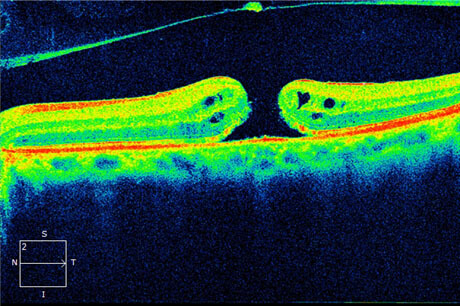
Pre-op Scan: Visual Acuity 20/400
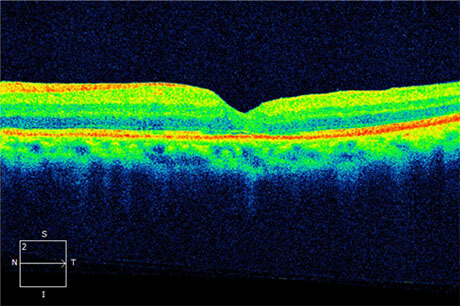
Post-op Scan: Visual Acuity 20/40
Patient 2:
Diagnosis: CRVO
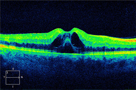
Pre-treatment Scan: Visual Acuity 20/60
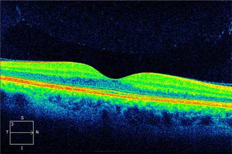
Post-treatment Scan: Visual Acuity 20/20
Fundus Scans
Patient 1:
Diagnosis: Central Retinal Vein Occlusion (CRVO)
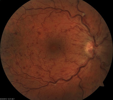
Pre-treatment Scan: Visual Acuity 20/60
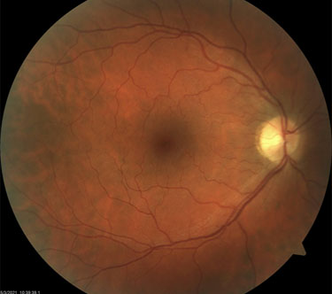
Post-treatment Scan: Visual Acuity 20/20
Patient 2:
Diagnosis: Branch Retinal Vein Occlusion (BRVO)
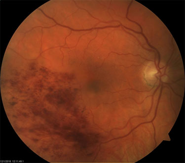
Pre-treatment Scan: Visual Acuity 20/60
Post-treatment Scan: Visual Acuity 20/20
Patient 3:
Diagnosis: Polypoidal Choroidal Vasculopathy
Pre-treatment Scan: Visual Acuity: Count Fingers
Post-treatment Scan: Visual Acuity 20/25
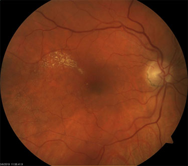
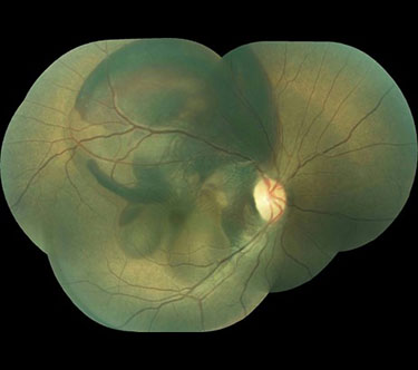
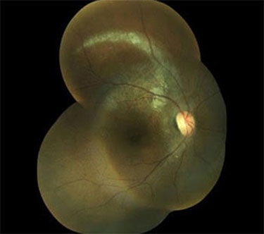
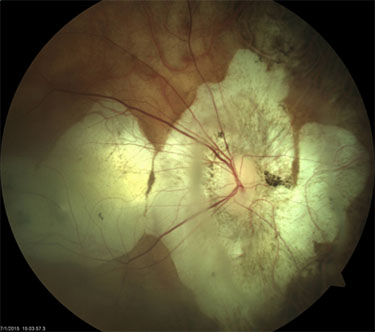
Fundus Scans Showing Other Different Pathologies

Wet Macular Degeneration
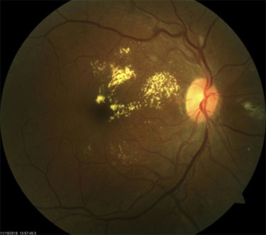
CSME
Myopic Degeneration
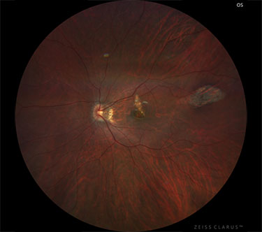
Multifocal Choroiditis ( MFC): Wide angle view
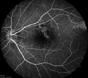
Multifocal Choroiditis (MFC): FA